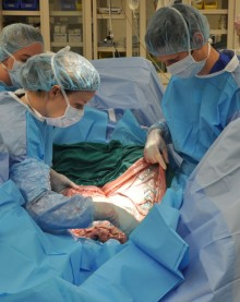
CALEC surgery, or cultivated autologous limbal epithelial cell transplantation, represents a groundbreaking advancement in the field of ophthalmology, offering renewed hope for patients with severe corneal damage. This innovative technique was successfully performed for the first time by Dr. Ula Jurkunas at Mass Eye and Ear, harnessing the body’s own stem cell therapy to restore the corneal surface. By sourcing limbal epithelial cells from a healthy eye, CALEC surgery can effectively treat eye injuries that previously left patients unable to regain their vision or alleviate pain. In a promising clinical trial, results indicated that over 90% of participants experienced significant improvement in their condition, showcasing the transformative potential of this procedure. As researchers continue to explore its implications, CALEC surgery could redefine eye injury treatment, providing life-changing outcomes for those affected by corneal damage.
Revolutionizing the treatment landscape for ocular trauma, the procedure known as CALEC surgery uses a patient’s own stem cells to mend corneal injuries that were once deemed irreparable. Often labeled as cultivated limbal epithelial cell therapy, this pioneering method involves harvesting healthy epithelial cells from the limbal region of the eye to combat the effects of severe corneal deficiencies. As clinical studies progress, particularly those conducted at prestigious institutions like Mass Eye and Ear, the focus on regenerating corneal surfaces has garnered significant attention. Notably, this approach stands to advance our understanding of stem cell applications in ocular health, paving the way for effective solutions in eye injury treatment. With ongoing research and trials, the future looks bright for patients suffering from the ramifications of corneal damage.
Understanding CALEC Surgery: A Breakthrough in Eye Therapy
CALEC surgery, or cultivated autologous limbal epithelial cell surgery, marks a significant advancement in the treatment of severe eye injuries, particularly for patients with corneal damage deemed irreparable. This innovative procedure utilizes limbal epithelial cells harvested from a healthy eye, which are then cultivated into a graft. Following a meticulous two to three-week manufacturing process at partner facilities, the graft is transplanted into the affected eye. This pioneering approach has demonstrated remarkable effectiveness, restoring the corneal surface in a majority of the patients involved in the clinical trials.
One of the key findings from the clinical trials at Mass Eye and Ear is that the CALEC procedure not only restores the integrity of the corneal surface but also significantly impacts patients’ quality of life by alleviating persistent pain and improving visual acuity. With over 90% success in restoring the corneal surface, the CALEC method offers new hope to individuals who previously had limited options for treating blinding corneal injuries. This success stems from the unique capability of limbal epithelial cells to regenerate the cornea, addressing the underlying issues caused by injuries such as chemical burns and trauma.
The Role of Stem Cell Therapy in Corneal Surface Restoration
Stem cell therapy represents a groundbreaking approach in the restoration of the corneal surface, particularly through innovative techniques like CALEC surgery. This therapy leverages the regenerative properties of limbal epithelial cells, which play a crucial role in maintaining the cornea’s structure and transparency. By cultivating these cells from a healthy eye and transplanting them into a damaged eye, researchers are pioneering a therapy that directly addresses the root of the problem rather than simply managing symptoms.
The clinical trial findings underscore the potential of stem cell therapy to change lives, as patients have shown marked improvements in both the physical state of their cornea and their overall vision. With a commitment to rigorous study and development, Mass Eye and Ear is leading the charge in establishing stem cell therapies as a viable and effective treatment option for those suffering from severe eye injuries that prevent conventional surgical options, such as corneal transplants.
The use of cultivated autologous limbal epithelial cells (CALEC) in the treatment process not only brings promise but also highlights the need for further trials to solidify its efficacy and deliver broader access to patients. It is an exciting time for ocular research, with stem cell therapy paving the way for future interventions that could transform eye injury treatment.
Eye Injury Treatment: Breakthrough Advances in Clinical Trials
The contemporary landscape of eye injury treatment is experiencing transformative changes, particularly with advancements presented in the recent clinical trials of CALEC surgery. Previously deemed irreversible, conditions such as limbal stem cell deficiency are now being approached with new methodologies that incorporate stem cell therapies. These innovative trials showcase a multifaceted approach to eye care, with teams like those at Mass Eye and Ear leading critical research in the application of advanced techniques to restore corneal integrity.
In these pioneering studies, clinicians are not only focusing on the physical restoration of the eye but also emphasizing patient well-being. The application of stem cell-derived treatments in clinical environments ensures that the latest research translates into tangible benefits for patients struggling with adverse effects of corneal injuries. Enhanced visual outcomes and quality of life improvements underline the importance of continued investment in clinical trials and the exploration of new treatment modalities.
The Future of CALEC Surgery in Eye Care
Looking ahead, the outlook for CALEC surgery and its role in eye care seems promising. As the research evolves, there is an ongoing interest in expanding the methodology to include an allogeneic manufacturing process which would utilize cadaveric donor eye tissue for cases where patients have injuries to both eyes. This innovation could potentially broaden the application of CALEC surgery and provide hope to a wider range of patients suffering from corneal deficiencies.
Collaboration among leading institutions and researchers continues to play a vital role in advancing these treatment approaches. As evidenced by the completed trials, which yielded promising results over the course of 18 months, future studies aim to include larger sample sizes and randomized control designs to further validate the effectiveness of CALEC surgery. Through meticulous research and innovation, we are on the cusp of redefining standards of care in ocular medicine, paving the way for a future where serious eye injuries can be effectively treated through regenerative therapies.
The Impact of Limbal Epithelial Cells in Eye Regeneration
Limbal epithelial cells are pivotal in the health and maintenance of the cornea, serving as the foundation for its regenerative capacity. These cells, found in the limbus of the eye, are crucial for repairing the corneal surface after injuries or disease. When these cells are compromised due to trauma, infections, or chemical burns, the ability to heal the cornea diminishes, leading to severe visual impairments. This is where the innovative CALEC procedure shines, as it reintegrates these essential cells into the cornea, promoting healing and recovery.
Research surrounding limbal epithelial cells has underscored their significance not only for eye health but also for broader applications in regenerative medicine. Through the painstaking process of harvesting and cultivating these cells, trials have demonstrated that stem cell therapy can effectively regenerate eye tissue. This not only restores vision but also opens avenues for further developments in treating various ocular diseases, positioning limbal epithelial cells as a cornerstone of future eye regeneration strategies.
Clinical Trials: The Backbone of CALEC Surgery Development
The successful development of CALEC surgery is intricately linked to the rigorous clinical trials conducted at Mass Eye and Ear. These trials not only provided critical insights into the safety and efficacy of the procedure but also validated the potential of stem cell therapy in treating severe corneal injuries. By carefully monitoring patient outcomes over time, researchers established promising metrics of success, including significant improvements in corneal surface restoration and vision.
These trials represent a collaborative effort across multiple disciplines, including ophthalmology and regenerative medicine, highlighting how interdisciplinary research can lead to breakthrough therapies. With ongoing support from institutions like the National Eye Institute, researchers are poised to expand upon these findings and explore new techniques that could further enhance the treatment of corneal injuries and expand therapeutic options available to patients.
Navigating the Regulatory Landscape for CALEC Therapy
Developing innovative treatments like CALEC therapy necessitates navigating a complex regulatory landscape to ensure safety and efficacy for patients. The trials at Mass Eye and Ear have adhered to the stringent guidelines set by authorities such as the FDA, allowing researchers to establish a robust framework for evaluating the application of stem cell therapies. Ensuring compliance not only reflects the commitment to patient safety but also lays down a pathway for potential approvals that could revolutionize ocular treatments.
As regulators continue to assess emerging therapies, public and private partnerships will be vital in pushing the envelope for CALEC surgery. Engaging with regulatory bodies early and often can facilitate the necessary discussions that move innovative treatments from exploratory stages to mainstream clinical use. This proactive approach could pave the way for CALEC therapy to become a widely accepted option for patients battling severe corneal injuries, ultimately enhancing the continuum of care within ophthalmology.
Patient Perspectives: The Human Side of CALEC Therapy
Patients are at the heart of every clinical trial, and their experiences provide invaluable insights into the effectiveness of new treatments like CALEC surgery. Many individuals who participated in the clinical trial reported significant improvements in their discomfort and vision, emphasizing the therapy’s emotional and practical benefits. The promise of restoring not just sight but also autonomy to those whose lives have been disrupted by debilitating eye injuries underscores the profound impact of this innovative procedure.
Moreover, the journey through clinical trials offers a profound narrative around hope and recovery. Patients who previously faced a bleak prognosis find renewed optimism as they embrace the possibility of treatment that can transform their lives. Delivering medical advancements with a clear understanding of patient experiences is crucial for further refining the techniques and ensuring that therapies not only restore vision but also enhance overall quality of life.
Collaborative Research Efforts in Ocular Stem Cell Therapy
The success of CALEC surgery can be attributed to the collaborative efforts across multiple research institutions. By pooling expertise from various fields, researchers are effectively driving innovation in stem cell therapy. This collaboration fosters an environment ripe for advancement, as teams at Mass Eye and Ear work alongside other institutions, including Dana-Farber, to refine techniques and share findings that push the boundaries of what’s possible in eye care.
The synergistic approach enables researchers to take advantage of diverse skill sets, accelerating the pace of discovery and application. By combining resources and knowledge, these partnerships are instrumental in studying the long-term effects of CALEC therapy and understanding its full potential in treating various ocular conditions. As such, the future of ocular health relies heavily on continuous collaboration in research, opening doors to exciting new possibilities for patients suffering from debilitating eye injuries.
Frequently Asked Questions
What is CALEC surgery and how does it work?
CALEC surgery, or cultivated autologous limbal epithelial cell surgery, is a groundbreaking procedure that transplant stem cells from a healthy eye to repair a damaged cornea. The process involves harvesting limbal epithelial cells from an unaffected eye, cultivating them in a lab to create a graft, and then surgically implanting this graft into the injured eye, effectively restoring the corneal surface.
What are the benefits of CALEC surgery for eye injury treatment?
CALEC surgery offers significant benefits for treating eye injuries that lead to corneal damage. Clinical trials have shown that CALEC has a success rate of over 90% for restoring the corneal surface, which is crucial for improving vision and reducing pain in patients suffering from severe injuries to their cornea.
How does CALEC surgery utilize stem cell therapy?
CALEC surgery employs advanced stem cell therapy by using cultivated stem cells—specifically limbal epithelial cells—from a healthy eye to repair the cornea of the injured eye. This innovative technique addresses limbal stem cell deficiency, which occurs when the cornea is damaged and can no longer regenerate the cells necessary for its surface.
What is the success rate of CALEC surgery in clinical trials?
In clinical trials led by researchers at Mass Eye and Ear, CALEC surgery demonstrated an impressive success rate, with 77% of patients experiencing complete restoration of their corneal surface at the 18-month follow-up. The results indicate that CALEC surgery is a highly effective option for patients with severe corneal damage.
Are there any risks associated with CALEC surgery?
CALEC surgery is generally considered safe, with no serious adverse events reported in clinical trials. Minor complications, such as a bacterial infection, occurred but were resolved without long-term effects. As with any surgical procedure, patients should discuss potential risks with their healthcare provider prior to surgery.
How is CALEC surgery different from traditional eye injury treatments?
Unlike traditional eye injury treatments, which may involve corneal transplants, CALEC surgery directly addresses the underlying issue of limbal stem cell deficiency. By using a patient’s own stem cells to regenerate the corneal surface, CALEC offers a targeted approach that can lead to improved outcomes and reduce the need for donor tissues.
Is CALEC surgery currently available for all patients?
Currently, CALEC surgery is experimental and not widely available. It is undergoing further clinical trials to establish its effectiveness and safety before it can receive federal approval for broader use in treating corneal injuries. Patients interested in this therapy should consult with eye care specialists at institutions conducting ongoing research.
What are limbal epithelial cells and why are they important in CALEC surgery?
Limbal epithelial cells are specialized stem cells located in the limbus of the eye, crucial for maintaining the cornea’s smooth surface. In CALEC surgery, these cells are harvested and cultured to create a graft that can replace damaged cells in the cornea, thereby restoring its functionality and alleviating symptoms of eye injuries.
What are the next steps for CALEC surgery research?
Future research on CALEC surgery aims to include larger clinical trials at multiple centers, longer follow-up periods, and randomized control designs. These studies will be essential to gather comprehensive data supporting the efficacy of CALEC and to facilitate its approval for broader clinical use.
How does the FDA relate to CALEC surgery?
CALEC surgery is under investigation as part of a clinical trial approved by the U.S. Food and Drug Administration (FDA). The FDA’s oversight ensures the safety and quality of the treatment as researchers gather evidence to support its potential approval for public use in treating corneal injuries.
| Key Point | Details |
|---|---|
| Introduction of CALEC surgery | Ula Jurkunas performs the first CALEC surgery at Mass Eye and Ear. |
| New hope for eye damage | Stem cell therapy shows promise for treating previously untreatable corneal injuries. |
| Effective treatment | Over 90% success rate in restoring corneal surfaces in clinical trials. |
| Stem cell process | Involves extracting stem cells from a healthy eye and transplanting them to a damaged one. |
| Clinical trial details | 14 patients followed for 18 months, showing significant cornea restoration. |
| Safety profile | No serious adverse events recorded during the trials. |
| Future research | Plans for allogeneic cell manufacturing to treat patients with bilateral damage. |
Summary
CALEC surgery represents a groundbreaking advancement in the treatment of corneal injuries, offering hope to patients previously deemed untreatable. This innovative procedure utilizes stem cells from a healthy eye to repair damage in an injured eye, achieving over 90% effectiveness in restoring corneal surfaces. The clinical trials have demonstrated a strong safety profile, with significant improvements noted in visual acuity among participants. As research continues, the potential for broader application and refinement of CALEC surgery highlights its role in transforming ophthalmic care and restoring vision.
